Transient Osteoporosis Of The Hip Mri

Transient Osteoporosis Of The Hip Radiology Reference Article Radiopaedia Org
Transient Osteoporosis Of The Hip Orthoinfo Aaos
Idiopathic Transient Osteoporosis Of The Hip Radiology Case Radiopaedia Org
Transient Osteoporosis Of The Hip Underlying Subchondral Fracture Radiology Case Radiopaedia Org

Transient Osteoporosis Not Just The Hip To Worry About Sciencedirect
Transient Osteoporosis Of The Hip Image Radiopaedia Org

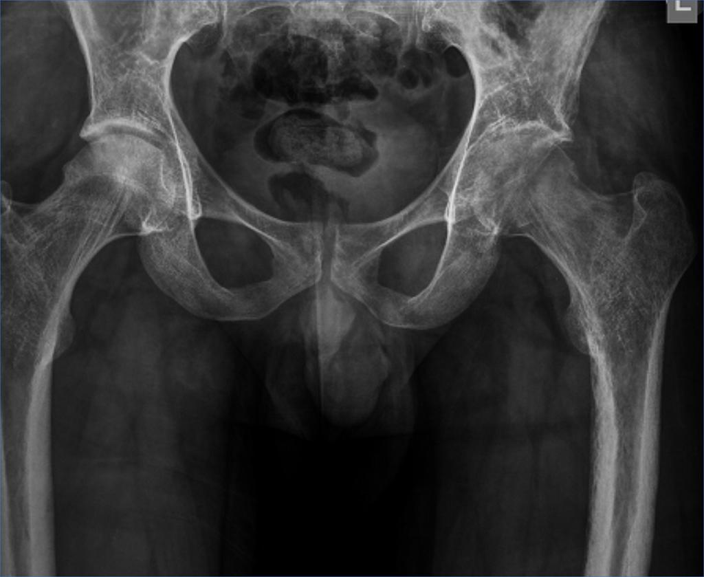
Toh was first described in 1959 in two women in their 3rd trimesters of pregnancy but now is more commonly seen in middle aged men.
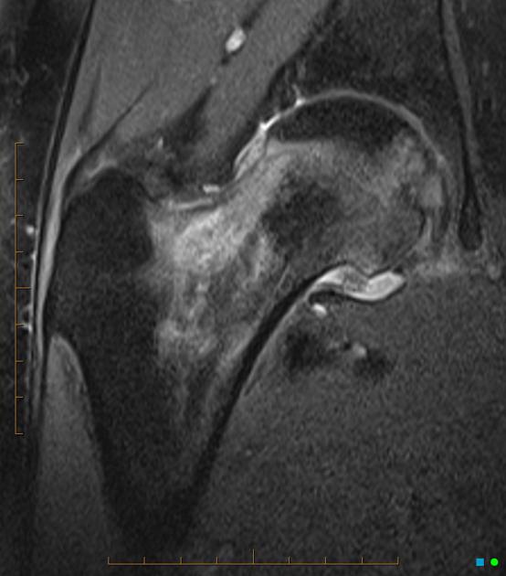
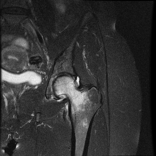
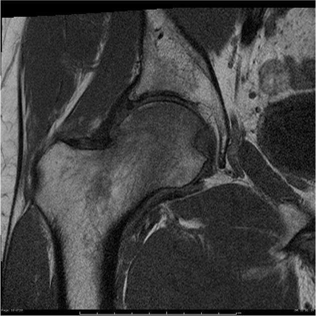
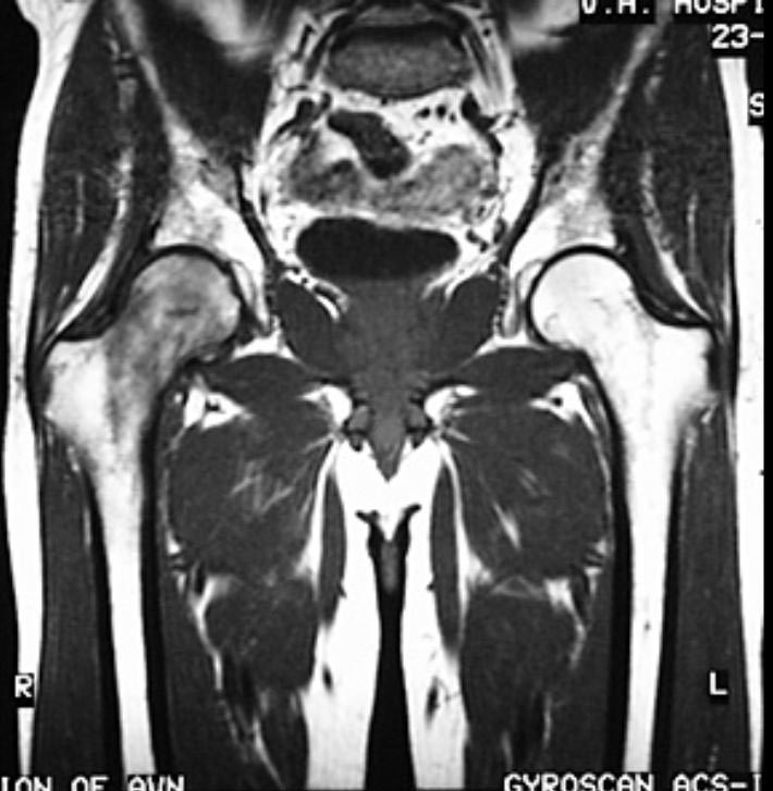
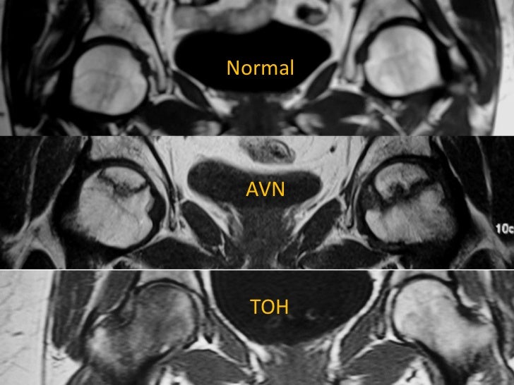
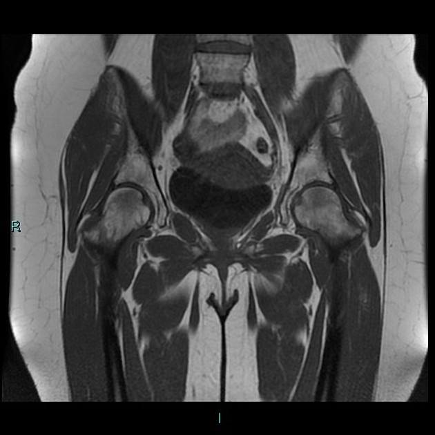
Transient osteoporosis of the hip mri. In this study we investigated the use of mri for evaluation of patients with transient osteoporosis to. Mri with t1 and t2 weighted sequences in coronal transverse and sagittal sections was performed in 12 patients with retrospectively confirmed to both at the onset of the disease and later as follow up procedure. By contrast mri changes in avn of the femoral head is restricted to a smaller. Mri changes in transient osteoporosis of the hip involve the whole femoral head and sometimes extend to the femoral neck and even the trochanteric region with blurred and vague margins.
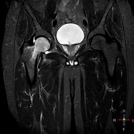
An mri scan of a hip affected by transient osteoporosis will usually reveal bone marrow edema. Three patients with transient osteoporosis of the hip underwent magnetic resonance mr imaging. This mri image shows edema surrounding the affected hip. Transient bone marrow oedema of the hip also referred as transient osteoporosis of the hip is self limited conditions that improves spontaneously over several months.
Mr images showed decreased signal intensity of bone marrow in the femur on t1 weighted images and increased signal intensity relative to the intensity of normal bone marrow on t2 weighted images.
Http Canjsurg Ca Wp Content Uploads 2014 03 46 3 187 Pdf
Idiopathic Transient Osteoporosis Of The Hip Radiology Case Radiopaedia Org
Https Www Journalofosteopathicmedicine Com Article S1746 0689 15 00133 9 Pdf
Http Www Akot Com Ar Cokiba Cursos 2018 16 Curso Oyt Files Transient 20osteoporosis 20of 20the 20hip 20review 20of 20the 20literature Pdf

Pdf Transient Osteoporosis Of The Hip
Transient Osteoporosis Of Hip

Annals Of Rehabilitation Medicine
Transient Hip Osteoporosis Radiology Case Radiopaedia Org

Transient Osteoporosis Of Pregnancy In A 34 Year Old Female Sciencedirect
Transient Osteoporosis Of The Hip Radiology Case Radiopaedia Org
Bilateral Transient Osteoporosis Of The Hip Postpartum Radiology Case Radiopaedia Org

Full Text Transient Osteoporosis Of The Hip Risk And Therapy Oarrr
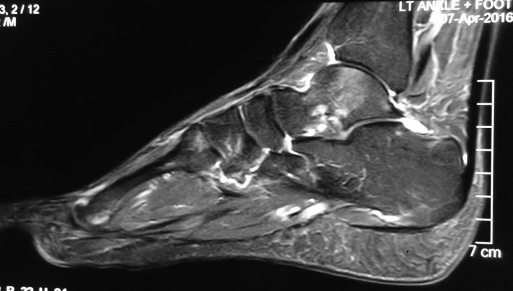
Transient Migratory Osteoporosis Of The Hip And Talus A Case Report Journal Of Orthopaedic Case Reports